
 L Lemo-Opuscar
显微镜故事
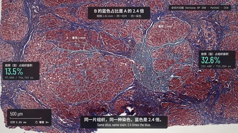
一镜从肝组织玻片推到单个细胞核,两个圆逐像素数出 2.4 倍的蓝。
- 适合:
- 一层层放大,讲眼睛、神经这类感知科普很合适
- 系列:
- L Lemo-Opuscar
- 更新:
- 2026-10-29
风格提示词
Microscope Story — Style Prompt
Real, licence-clean microscopy of a real specimen, seen as it is, and one continuous change of scale that carries the viewer from the glass slide in the hand to a single cell. Focus breathes when the magnification changes, a focal plane sweeps when the camera lands, and every scale bar, field of view and reading comes from the image's own metadata.
References (grammar only): Charles and Ray Eames' Powers of Ten (one continuous change of scale with a readout riding along); premium microscope launch films (dark bench, glass catching light, restrained marks, clean instrument sound); digital-pathology viewers' instruments (scale bar, field of view, magnification). Copy none of their layouts, interfaces, typefaces or music; never name them in the film.
1. Essence, and what it is not
- The specimen is a photograph: a real micrograph or whole-slide scan of the actual sample, CC0, public domain or CC BY. Nothing on the specimen is drawn, retouched or generated. A whole-slide image (WSI) is the ideal source: one sample, one registered pyramid from millimetres to microns, with the pixel size in its metadata.
- Scale is the camera: one log-space dive (and, when the story wants it, the climb back) through nested plates of the same sample. The scale bar shortens and jumps in a 1-2-5 sequence, the field-of-view readout rolls, the magnification steps.
- Focus has a grammar: a rack-in from soft at the start, a short breath (blur + a hint of field change) each time the magnification changes, a focal-plane sweep across the frame when the camera lands.
- Readings are measured: every number is computed from the file (pixel size × pixels) and the screen says how it was measured. Describe what was measured; never diagnose. Keep a
FACTS.mdin the project folder (claim, source URL, quoted wording, or the measurement method) and deliver it with the film. - One reading per screen, in the single accent colour.
Not Satellite Briefing (no grey grade; the stain keeps its colour), not Sci-fi HUD (no glowing wireframes or crosshairs), not Data Storytelling (no chart paper), not a keynote (no slides or header bars).
2. Materials & rendering
- Plates: the WSI pyramid levels as plates in one world frame (level-0 pixels), the coarse levels whole and alpha-feathered at the scan edge, the fine levels as crops around where the camera lands. Each frame draws the finest plate that is downsampled no more than ~1.45×, and blends the next level in between 2.2× and 1.45×: no seams and no visible upsampling, because every level is the same sample, already registered.
- The naked-eye plate: the scanner's own photo of the whole slide (many WSI files carry one), registered through the scan's area-of-interest metadata and checked against the tissue's centroid. Cut the glass out of the scanner tray, remove the line-sensor streaks, and fill any manufacturer's printed logo with the surrounding frosted glass. Below ~30 mm of field the photo would be upsampled: let the cover glass become clean glass (its dust leaves the focal plane).
- One grade for every plate: glass normalised to a warm white, a little density and chroma in the stain, nothing else. No false colour, no film emulation.
- The bench: graphite under a slow pool of light; a soft shadow under the slide.
- Focus: screen-space Gaussian blur on a frame rendered with a margin (so edges never go dark); the focal sweep is a tilted sharp front crossing a softened frame. A light illumination vignette, nothing more.
- Marks in screen space but anchored to the specimen through one world → screen function: hairlines 1.5–2.5 px with a dark or light halo, circles drawn on as arcs with cardinal ticks, dimension lines with end ticks, leader lines with one elbow. Counted pixels are shown as a mask revealed behind a scan front, then kept as a faint trace.
- Frosted panels (dark translucent glass over a blurred copy of the frame) for readings, instruments, subtitles and the credit: white type reads on glass and stain alike, nothing switches colour mid-shot.
3. Colour logic
- Glass: warm paper white. Bench: graphite. The stain is evidence and keeps its true colour (e.g. trichrome red and blue, H&E pink and purple).
- One accent, cool and away from the stain's hues (mint against red and blue; amber against a green or purple stain), only for the measured reading and the counted pixels.
- Ink on glass is near-black; type on stain sits on a frosted panel in warm white.
Example (our demo): glass #F4F1EC, bench #0B0D0F → #2A3036, ink #181B1F, panel rgba(12,14,17,0.74), accent mint rgb(120,240,205) (on stain) / rgb(0,122,99) (on glass).
4. Type & subtitles
- Roles: a light condensed grotesk for readings and the scale bar (40–72 px, e.g. Barlow Condensed); a monospace for instruments and pixel counts (20 px, e.g. IBM Plex Mono); a sans in the film's language for labels (20–34 px, e.g. Noto Sans SC); a serif only for the title. OFL, self-hosted, CJK subset.
- Instruments live in one frosted panel in a corner: scale bar with its value, field of view, magnification with a small nosepiece glyph that turns a quarter on every change. The source credit in a small panel in the opposite corner.
- Readings sit in a frosted panel beside what they measure, joined by a leader: a small label above ("collagen share of tissue area"), the number large in the accent, the method or the raw counts below.
- Subtitles: a frosted strip at the bottom; if the film is bilingual, one line per language with the film's language on top, never wrapped. Measure each line with the real font and split long lines into two cues.
- Nothing below 20 px at 1080p. Hold labels ≥ characters ÷ 4.5 + 1.5 s (CJK); a reading that rolls is reported by its landing value.
5. Motion quality
- The camera is a similarity transform (zoom, roll, position) interpolated as a log spiral between keys (M^u about the fixed point of the two frames), so every push is a true dive towards one point and the roll is part of it. Ease the moves, creep 2–5 % during holds; never a frozen frame longer than a second.
- Magnification changes happen where the plates actually change, each with a breath of focus, a turret click and the new magnification said once, large.
- Marks arrive by being made: a dimension line grows from its centre, a circle is drawn as an arc, a scan front counts pixels while the reading rolls, the reading lands with a small spring.
- Never moves: the specimen under its marks, the instrument positions.
6. Camera grammar
| Move | What it expresses | Can serve |
|---|---|---|
| The dive (log zoom with roll) | going to look for yourself | an opening; from the slide to the cell |
| Locked frame, slow creep | evidence, a measurement | a reading; a comparison |
| Truck at one magnification | a second sample of the same thing | the counter-example; "meanwhile, nearby" |
| Pull back to frame two marks | comparison | the verdict |
| Focal-plane sweep | arriving; depth | a landing; a reveal inside one frame |
| The climb (reverse dive, scale bar jumping back) | consequence, scale | an ending; a return to the eye's scale |
At key moments the measured structure fills a third of the frame height or more; instruments in a corner, subtitles bottom centre, readings beside their marks. Transitions are scale changes, trucks and focus, never cuts or crossfades between samples.
7. Sound palette
- Instrument foley: the objective turret (a click, a detent, a small metal ring), a focus servo, pen on glass for each line drawn, a band-passed scan travelling left to right with counter ticks that speed up, a soft filtered sweep for the focal plane.
- Music: hard-mallet vibraphone in 8ths and 16ths, marimba ticks, bowed vibraphone and water glasses for pads and the motif, glockenspiel for each reading that lands, a pulse bass with 2nd/3rd harmonics, timpani and bass drum under the landings. Harmony stays open and light; the score is the user's to shape.
- Beds: a quiet lab room (fan, a little mains hum).
- Silence before the verdict and before the climb; the first sound after it is the reading or the turn.
- Mix: voice ~10 dB over music, music ducked under it; −14 LUFS. Voice: a calm, exact narrator; numbers spelled out for the TTS, digits on screen.
8. Native moves
- The dive. From the slide in the hand to a cell in one camera, the turret clicking at each real change of level. Fits content like: a biopsy to its cells; a coated surface to its crystals; a hair or skin section to its layers.
- Counting the colour. Draw a circle, let a scan front cross it, light up every counted pixel and roll the share up to its landing value; show the raw counts underneath. Fits content like: collagen in tissue; porosity in a ceramic; coverage of a coating; pigment in a sample.
- Two circles, one verdict. Measure one place, truck to another at the same scale, then frame both and say what differs. Fits content like: treated against untreated; edge against centre; before against after.
- The focal sweep and the ring. Land, sweep the focal plane across the frame, ring one structure and give its size. Fits content like: a nucleus; a grain; a fibre's diameter.
- The climb back. Pull out to the eye's scale with the scale bar jumping backwards; a title card written on the glass.
9. Pitfalls of the medium
- Licences: most microscopy on the web is CC BY-NC or all rights reserved. Usable: CC0 whole-slide scans (e.g. the OpenSlide test data: check each file's licence line), public-domain government micrographs (NIH, CDC PHIL, NASA, USGS), CC BY collections (Cell Image Library, Wellcome Collection), with every file listed in
CREDITS. Not usable: CC BY-SA without agreement, NC images, journal figures, AI images. - Finding material for a new topic: search by organ, stain or material plus "whole slide" in the OpenSlide test data, NCI Imaging Data Commons (TCGA/CPTAC slides, licence per collection), the Cell Image Library, Wellcome Collection, Wikimedia Commons (filter by licence) and NIH/CDC galleries; prefer a file whose metadata carries its pixel size. CC BY-SA, ODbL or "no known restrictions" are not CC0: name the terms to the user and ask before using them. For a product, a lab or a brand, ask the user for their own scans (SVS/NDPI/MRXS, SEM TIFFs with a scale) first. Never stand in a generated image for a real specimen.
- Different samples never zoom into each other: two micrographs of "the same thing" at two scales are two specimens. Use one pyramid; if you must join sources, cut on a focus breath and say so.
- Pixel size: read it from the file (XMP
ScanRes, TIFF resolution, OMEPhysicalSizeX); a slide-photo's resolution tag can be trusted only after checking against the scan. - Upsampling shows as mush beyond ~1.6×: end the dive at the scan's real resolution.
- Tile seams: some scanners overlap tiles; check the joint info, frame out visible seams.
- Labels and logos on the slide: barcodes and IDs are part of the object; manufacturer logos are not — fill or crop them.
- Claims: a stain's colour means what the stain protocol says, nothing more; measured shares are of the drawn region and the stated threshold, never a clinical score.
10. Engine
demo/film.js: log-spiral camera (camAt), plate selection by downsampling ratio and coverage, objective events from real level changes, focus, frostedpanel(), marks, readings,TEXTS/SUBCHECK/EV.demo/tools/plates.py: slide-photo registration and cleaning, graded plates, collagen mask and the scan's cumulative curve, the tissue axis.nucleus.py: membrane edge on 72 rays, least-squares circle.- Extra packages and packs (beyond
setup.sh deps voice):tifffile+imagecodecsread TIFF-based slides (Ventana BIF, Aperio SVS, Hamamatsu NDPI, generic pyramidal TIFF;demo/tools/plates.pyis written for BIF: its label page, XMP and AOI); MRXS, SCN, VMS and others needopenslide-pythonwith the OpenSlide library (brew install openslide), or an adapter of your own. Install them yourself into a private venv in the project folder, not into$LIB/.venv. - Minimal example, not in the demo: a coated steel sample: one SEM pyramid, a dive to one crack, a circle counting coverage, a ring around one grain.
11. Variation space
The agent decides the specimen, the stain or modality, what is counted, the camera path, the accent, the ending.
- Structures: a single dive ending in one measurement; a comparison of two regions or two samples at one scale; a climb that starts on a cell and reveals the object.
- Openings: the slide on the bench under moving light; a cell already in focus, the climb revealing what it belongs to; a blurred field racking into focus.
- Endings: the title written on the glass; a last reading held in silence; the slide lifted out of frame.
How our demo was made (story, shots, score, title card, build): DEMO.md. Read it after your treatment exists.
补充
示范片
Measured Blue
本风格的来源项目:开源项目 Lemo-Opuscar(项目仓库)。本站保留原项目署名,具体许可以该项目仓库为准。
分享来源:B 站视频(341 种代码动画风格)